NEURORADIOLOGY พ.อ.หญ ง อนงค ร ตน เกาะสมบ ต กองร งส กรรม โรงพยาบาลพระมงก ฎเกล า
|
|
|
- Shannon Gregory
- 5 years ago
- Views:
Transcription
1 NEURORADIOLOGY พ.อ.หญ ง อนงค ร ตน เกาะสมบ ต กองร งส กรรม โรงพยาบาลพระมงก ฎเกล า
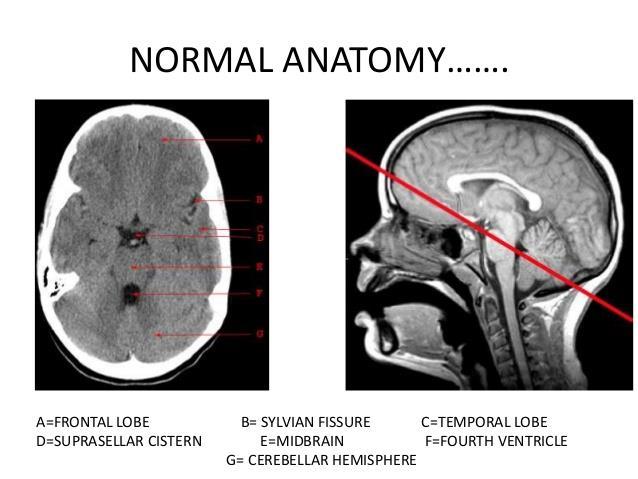
2 CONTENTS Modality and indication, limitation, contraindication Plain film positioning Normal and abnormal plain film Emergency CT of the brain Clinical application
3 IMAGING MODALITY Plain film Computer tomography Cerebral angiography Magnetic resonance imaging Ultrasound
4 PLAIN FILM ศ กษาความผ ดปกต ในการเจร ญของศ รษะและใบหน า ด ความผ ดปกต ของกระด กจากการทาลายเช น infection, metastatic tumor ด fracture หร อ foreign body ใช เป นหล กฐานทาง forensic medicine
5 Computer tomography Indication Head injury Stroke Increased intracranial pressure - Tumor, hydrocephalus,abscess Pre-op or follow up after surgery
6 Computer tomography Limitation Bone artifact Isodense lesion Radiation hazard Contrast media risk
7 Cerebral angiography Indication abnormal vessels(aneurysm,avm) Tumor vascularity, pre-op evaluation Vasculitis Carotid cavernous fistula, pseudoaneurysm
8 Cerebral angiography Advantage Hemodynamic study, flow and direction Intervention Disadvantage Invasive Contrast media Radiation
9 Magnetic resonance imaging Indication Tumor Stroke Perfusion study Angiogram : MRA Specific areas: pituitary, IAC, cranial nerves,orbits
10 Magnetic resonance imaging Advantage High tissue characteristics and resolution Multiplanar imaging No radiation Limitation Long acquisition time Poor bone and calcium details High cost Claustophobia
11 Magnetic resonance imaging Contraindication Clip aneurysm Pacemaker Ferromagnetic substance Metallic foreign body Long term hazard
12 Ultrasound Infant brain ผ านทาง fontanelle, anterolateral, posterolateral fontanelle Color doppler US for carotid or vertebral arteries
13
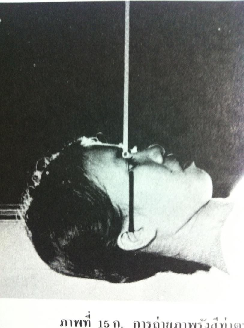
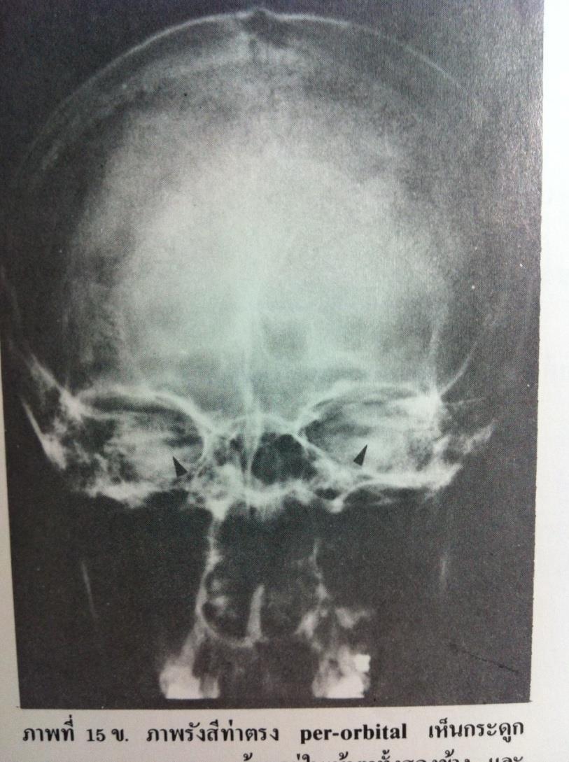
14 ORBITOMEATAL LINE The line passes through the outer canthus of the eye and the center of the external auditory meatus. It is used for positioning for radiographic views including Water's view, Perorbital view, Lateral view, and others.

15 LATERAL VIEW

16 CALDWELL S VIEW
17 AP VIEW

18 TOWNE S VIEW

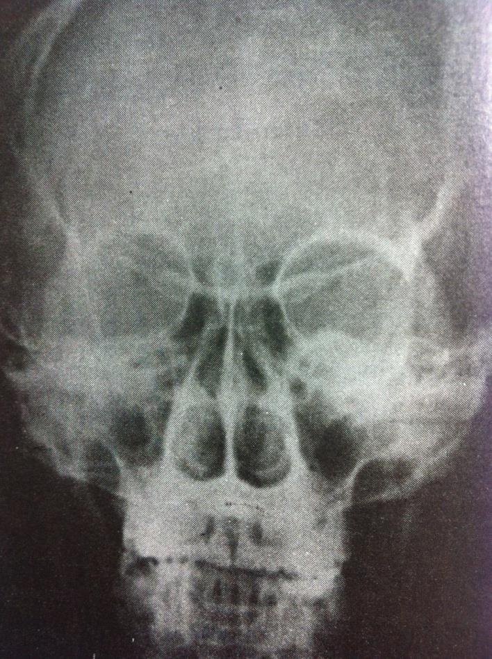
19 WATER S VIEW

20 NORMAL CALDWELL VIEW Inner,outer table Frontal sinus Cribiform plate Ethmoid sinus SOF Petrous apex

21 NORMAL LATERAL VIEW Coronal,lambdoid,squamosal MMA groove Arachnoid granulation Sphenoid sinus mastoid air cells

22 NORMAL TOWNE S VIEW Occipital bone Petrous, IAC Lambdoid suture Transverse sinus Foramen magnum Mandibular rami Zygomatic arch
23 NORMAL WATER S VIEW Maxillary sinus Zygomatic arch Mandible Maxilla Nasal cavity Orbit

24 CONED SELLA VIEW Anterior clinoid process Posterior clinoid process Sella turcica Dorsum sellae Tuberculum sellae
25 หล กการแปลผล PLAIN FILM SKULL Size and shape: 1:4-1:1.5 Cranial vault : Thickness, symmetry, sutures,convolutional markings, vascular groove Skull base: Sella turcica Calcification: Facial bones: orbits, PNS, mandible, mastoid, maxilla Soft tissue
26 Types of fractures Linear fracture Diastatic fracture Comminuted fracture Compound fracture Depressed fracture Base of skull fracture: CSF rhinorrhea Fracture via sinus, mastoid: pneumocephalus
27 PHYSIOLOGIC CALCIFICATION Pineal gland: 2/3 adult,less than 10mm Habenula : anterior to pineal gland, C shape Choroid plexus: Atrial of lateral ventricles Basal ganglia: Bilateral punctate or conglomerate. Idiopathic, Fahr disease, CO or lead poisoning, metabolic PTH, mitochondrial disease, MELAS Dura: Falx cerebri,tentorial cerebelli,petroclinoid ligament
28 PATHOLOGIC CALCIFICATION Chronic SDH, old hematoma Parasite Infection: TB, abscess CMV, Toxoplasmosis Vascular: Atherosclerosis,AVM Tumor: Oligodendroglioma, Meningioma, Craniopharyngioma
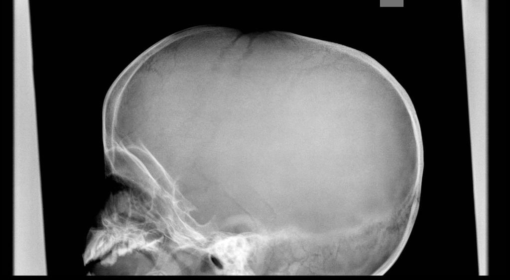
29 Increase intracranial pressure Enlarged cranial cavity Convolutional marking Suture separation Erosion of dorsum sellae
30 Enlarged pituitary fossa Ballooning/ J-shape sella Pituitary gland: adenoma, Rathke cleft cyst Parasellar structure: Meningioma, aneurysm Suprasellar structure: Craniopharyngioma, weakness of diaphragmatic sella, arachnoid cyst

31 J-Shaped sella
32 Sclerotic skull lesions Osteoblastic metastasis Paget s disease, osteopetrosis, fibrous dysplasia Fluorosis Chronic anemia: Diploic space,hair on end periosteal reaction Acromegaly: Thick skull, enlarged sinus and mastoid, sellar enlargement
33 Osteolytic skull lesions Congenital : meningoencephalocele Trauma: leptomeningeal cyst Infection: Osteomyelitis Tumor: epidermoid, Hisiocytosis X, hemangioma Miscelleneous: Paget, FD, hyperpth
34 BASIC CT BRAIN INTERPRETATION

35 What is a CAT scan? Cross sectional images are obtained by multiple measurements of the x-rays attenuation from several projections.
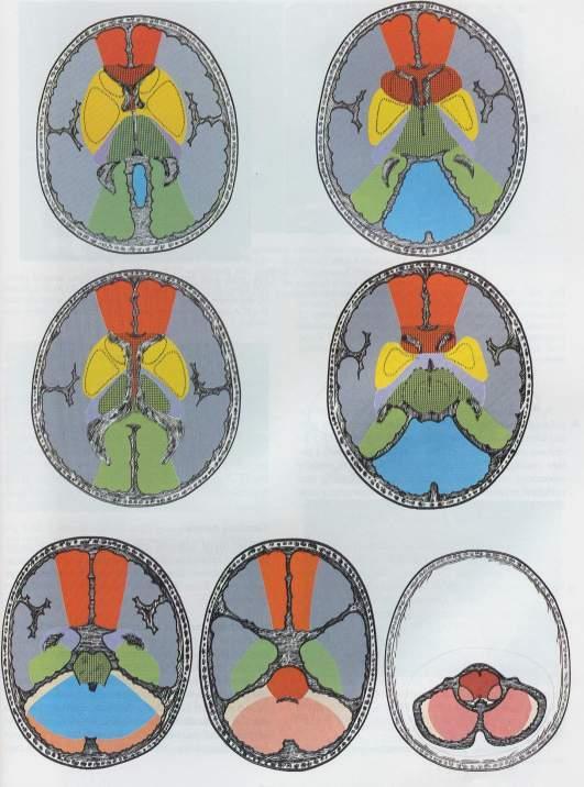
36 SYSTEMATIC APPROACH Symmetry Cross sectional anatomy Window Brain Blood Bone
37

38
39
40
41
42
43
44
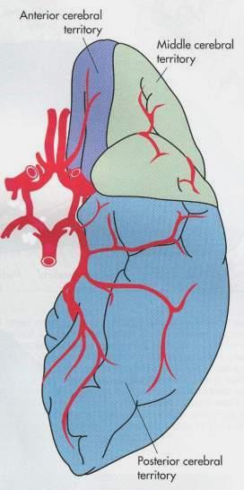
45 Arterial Territories Anterior Cerebral Middle Cerebral Posterior Cerebral Basilar Superior Cerebellar Anterior Inferior Cerebellar Posterior Inferior Cerebellar
46 Supratentorial Territories

47 From Osborne, A: Neuroradiology
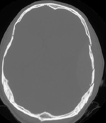
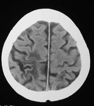
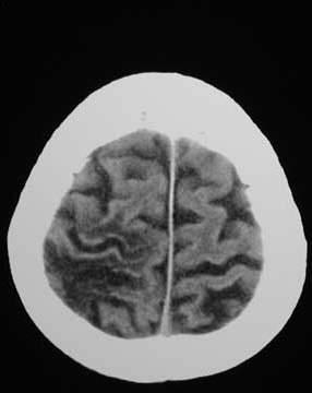
48 EMERGENCY NEURORADIOLOGY Plain film: Limit evaluation of intracranial pathology CT: Fast, accessible, unstable patient Brain, bone, blood window Limitation: beam hardening artifact at posterior fossa
49 TRAUMA - Primary effect : Contusion, hemorrhage - Secondary effect : Herniation syndrome, vascular injury and its complication
50 EXTRAAXIAL HEMORRHAGE Epidural hematoma Subdural hematoma Subarachnoid hemorrhage
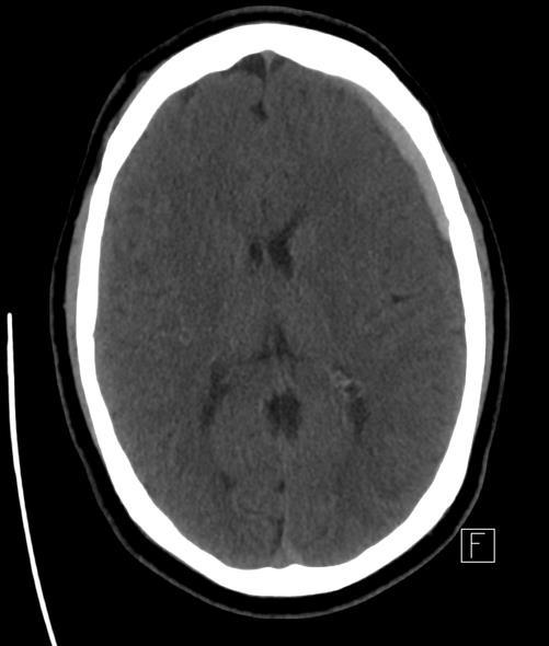
51 EPIDURAL HEMATOMA Bleeding between inner table of skull and outer layer of dura Cause : arterial (90%), venous, other Location : Squamous portion of temporal lobe which contain middle meningeal artery Not cross suture line Biconvex hyperdensity lesion Associated skull fracture common

52 Epidural Hematoma
53 SUBDURAL HEMATOMA Acute Subacute Chronic : Uncomplicated, complicated, mixed (acute on top)
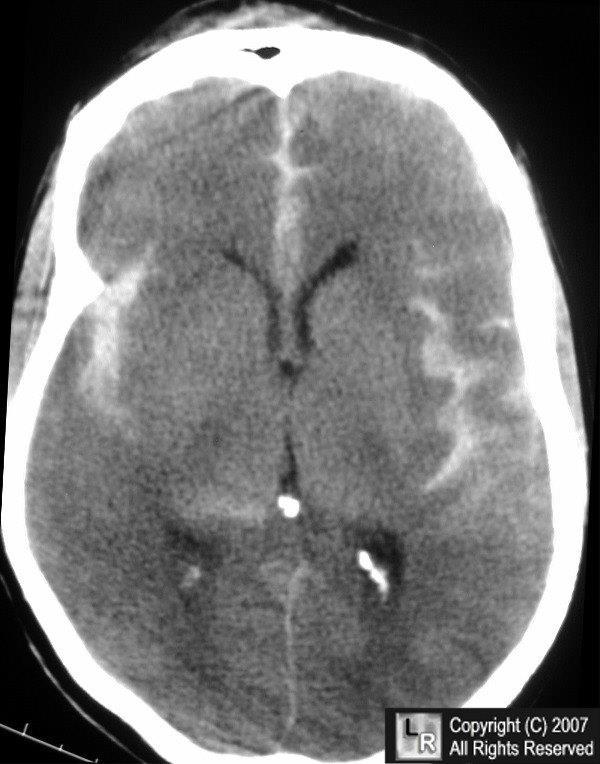
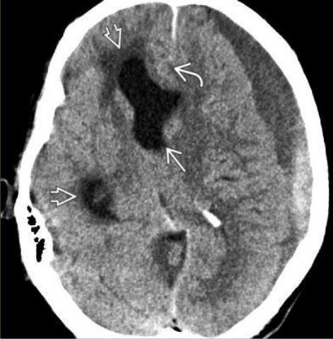
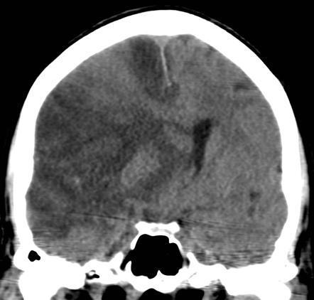
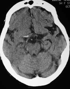
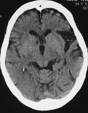
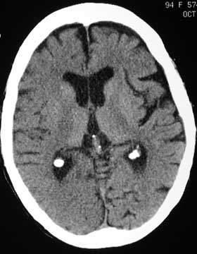
54 SUBDURAL HEMATOMA Bleeding between inner layer of dura and arachnoid Cause : tearing of cortical vein is most common, artery, aneurysm, metastasis, spontaneous, etc Can cross suture line, not cross dural reflection (falx cerebri and tentorial cerebelli)
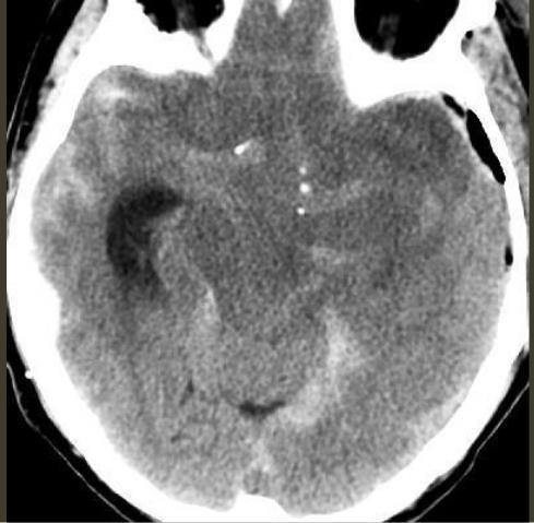
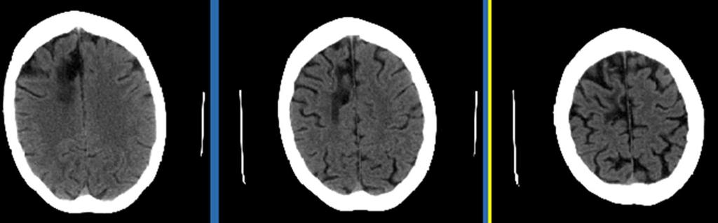
55 SUBDURAL HEMATOMA Acute : Crescent shape hyperdensity along hemispheric convexity Subacute : Isodensity lesion to brain parenchyma (density decrease 1.5H.U. per day) Bilateral subacute SDH hematoma: - may be undiagnosed - Sulcal effacement and medially displaced gray-white interface
56 Subdural hemorrhage

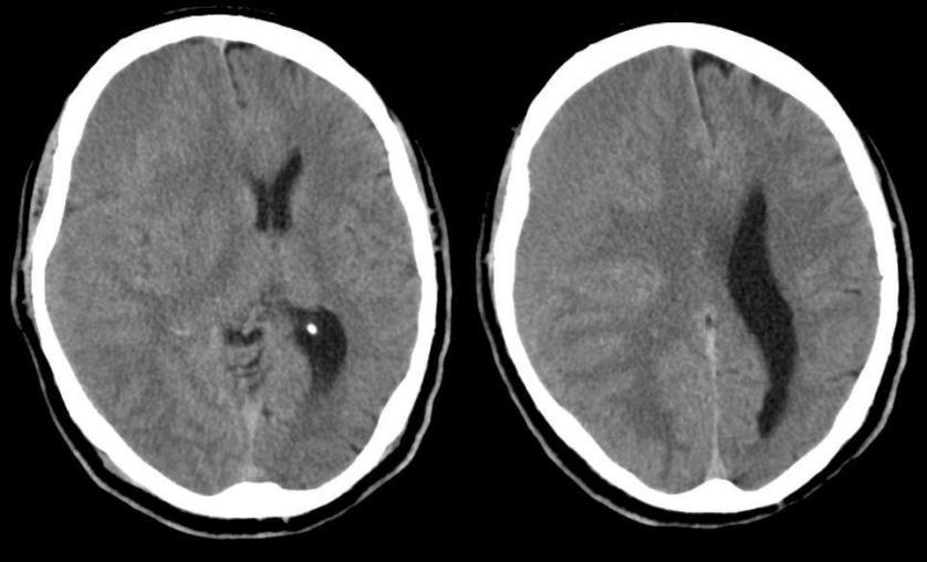
57 Subacute subdural hemorrhage
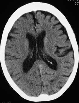
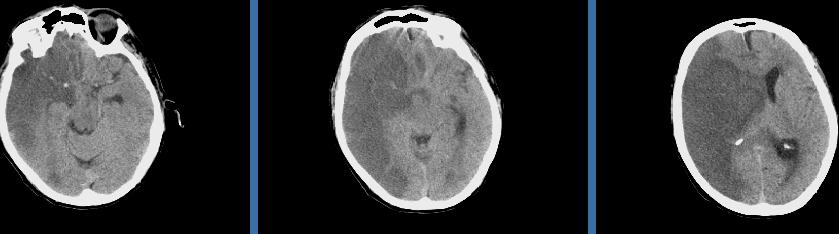
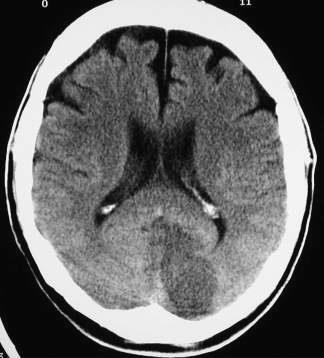
58 SUBDURAL HEMATOMA Chronic : Hypodensity, near CSF density in uncomplicated case Complicated chronic subdural hematoma : Thick membrane, internal septation, loculation from repeated hemorrhage Mixed subdural hematoma : Rebleed into the chronic subdural hematoma, (Acute on top) Hematocrit level

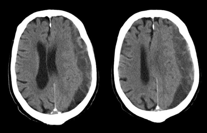
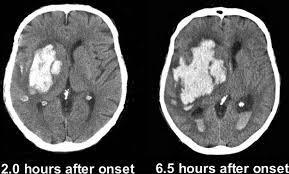
59 Chronic subdural hemorrhage

60 Mixed subdural hemorrhgae
61 SUBARACHNOID HEMORRHAGE Bleeding between arachnoid and pia mater Mild irregular linear hyperdensity in the cistern and sulci Location : Sylvian fissure, cortical sulci, suprasellar cistern, interpeduncular cistern, basal cistern Cause -Traumatic - Non traumatic : aneurysm, AVM
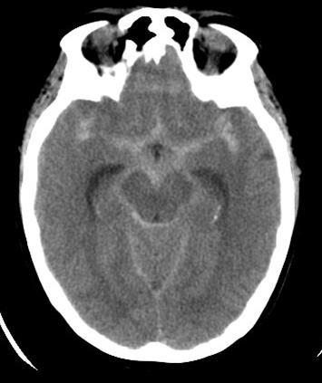
62 SUBARACHNOID HEMORRHAGE
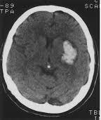
63 INTRACEREBRAL HEMORRHAGE Cerebral contusion Intraparenchymal hemorrhage - Traumatic - Non traumatic
64 CEREBRAL CONTUSION Brain bruise, impacted against bony ridge or falx cerebri Depressed skull fracture Location: temporal lobe, anteroinferior frontal lobe CT: Hypodensity from cerebral edema with petechial hemorrhage
65 INTRACEREBRAL HEMORRHAGE CAUSE Benign: Hypertension, vascular lesion, trauma, drug, coagulopathy Malignant: Primary or metastatic tumor
66 INTRACEREBRAL HEMORRHAGE Benign findings: Round to oval shape hyperdensity lesion Swirl sign : inhomogeneous density due to active bleed Malignant findings: Soft tissue density Variable age of blood Vasogenic edema inappropriate to lesion No evolution with time
67 Hypertensive hemorrhage Cause : Atherosclerosis lentriculostriate artery most affected Ruptured Charcot Bouchard aneurysm Common location Basal ganglia, thalamus, brain stem and cerebellum. Lobar hemorrhage 5% CT findings Homogeneous round to oval shape hyperdensity in typical location. May have intraventricular extension
68
69 HERNIATION SYNDROME 1. Subfalcine herniation 2. Transtentorial herniation: ascending, descending 3. Tonsillar herniation 4. Transalar herniation
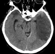
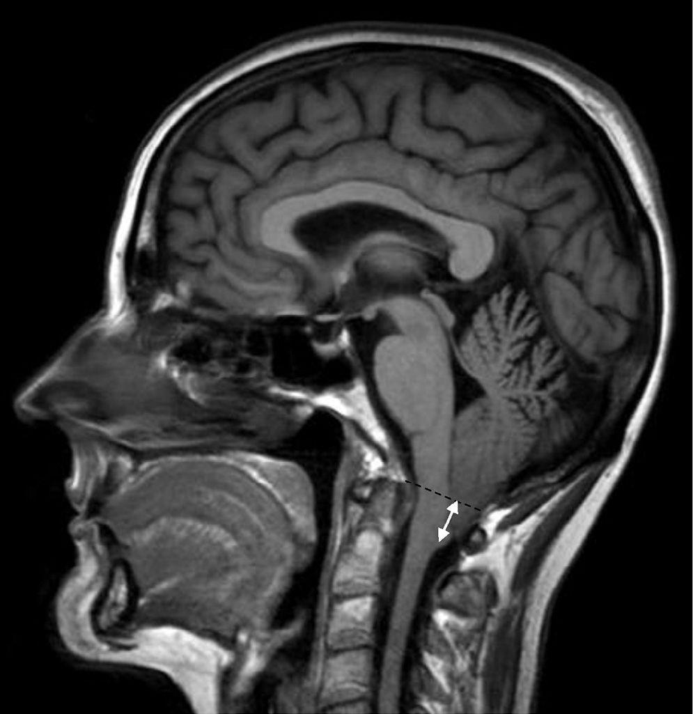
70 SUBFALCINE HERNIATION 1.Herniation of affected hemispheric structures across midline under the free margin of falx cerebri 2.Involve corpus callosum, cingulate gyrus, anterior cerebral artery and internal cerebral vein 3.Complication : - Obstructive hydrocephalus - ACA infarction of cingulate gyrus
71 SUBFALCINE HERNIATION 4.Imaging : - Shift of midline structures - compressed ipsilateral ventricle - obstructive hydrocephalus with dilatation of contralateral ventricle - Hypodensity of cingulate gyrus due to ACA infarction.
72
73 TRANSTENTORIAL HERNIATION Descending transtentorial herniation 1. Herniation through tentorial incisura 2. Involve uncus, hippocampus, brain stem, cranial nerve III, PCA 3. Complication - Third nerve compression and palsy - PCA occlusion and infarction - Kernohan notch - Duret hemorrhage
74 TRANSTENTORIAL HERNIATION Descending transtentorial herniation 4.Imaging : - Medial displacement of uncus of tempotal lobe - Effacement of ipsilateral aspect of suprasellar cistern - Shifting of midbrain against the contralateral tentorial ridge - Hypodensity of contralateral cerebral peduncle from ischemic injury (Kernohan notch). False localizing sign - Duret hemorrhage due to compressed perforator of basilar artery - PCA infarction; Hypodensity of occipital lobe

75 Ipsilateral suprasellar cistern effacement
76
77 TONSILLAR HERNIATION Herniation of cerebellar tonsil via foramen magnum Complication: Obstructive hydrocephalus Imaging : CT : Obliteration of CSF spaces around in cisterna magna MRI Sagittal view: tonsil >5mm below the foramen magnum
78 Tonsillar herniation
79 Ascending transtentorial herniation Less common Upward herniation of cerebellar through tentorial incisura Cause : Posterior fossa mass lesion. Complication: Obstructive hydrocephalus Imaging : CT : Obliteration of superior vermian cistern and quadrigeminal plate cistern.

80 Ascending transtentorial herniation
81 STROKE - Ischemic stroke - Hemorrhagic stroke
82 Ischaemic Stroke Thromboembolic most common Hemodynamic Atherosclerotic Dissection Vasospasm Hypotensive /asphyxia (watershed) Migraine Vasculitis Thrombotic: hypercoagulable states
83 Hemorrhagic Stroke Primary Intracerebral hemorrhage Hypertension Amyloid angiopathy Arteriovenous malformations Neoplasms Trauma Subarachnoid hemorrhage Aneurysm AVM s Trauma
84 Hyperdense vessel sign & loss of gray/white junction...


85 Left insular ribbon sign & effacement of sulci
86 ACA MCA


87 Left PCA

")
88 Watershed (between ACA and MCA)
89 Hypertensive Hemorrhage Classically involves the deep nucleii
90 INTRACRANIAL INFECTION Intraaxial: Encephalitis Cerebritis Abscess Extraaxial: Subdural empyema Epidural abscess Meningitis
91 CEREBRAL INFECTION Encephalitis: generalized and difuse infection of the brain. Often of viral origin (ex.herpes simplex) Cerebritis: localized but poorly demarcated area of parenchymal softening. Abscess: follows cerebritis. Occurs when a central zone of necrosis becomes encapsulated.
92 MODE OF SPREAD Hematogenous spread Direct extension: ex.sinusitis leading to epidural abscess or subdural empyemas Spread along the nerves (ex.herpes encephalitis along the trigeminal nerve)


93 SUBDURAL EMPYEMA (C+)

94 TUBERCULOUS MENINGITIS Basal leptomeningitis (seeding of the subarachnoid space)

95
NEURO IMAGING 2. Dr. Said Huwaijah Chairman of radiology Dep, Damascus Univercity
 NEURO IMAGING 2 Dr. Said Huwaijah Chairman of radiology Dep, Damascus Univercity I. EPIDURAL HEMATOMA (EDH) LOCATION Seventy to seventy-five percent occur in temporoparietal region. CAUSE Most likely caused
Head CT Scan Interpretation: A Five-Step Approach to Seeing Inside the Head Lawrence B. Stack, MD
 Head CT Scan Interpretation: A Five-Step Approach to Seeing Inside the Head Lawrence B. Stack, MD Five Step Approach 1. Adequate study 2. Bone windows 3. Ventricles 4. Quadrigeminal cistern 5. Parenchyma
For Emergency Doctors. Dr Suzanne Smallbane November 2011
 For Emergency Doctors Dr Suzanne Smallbane November 2011 A: Orbit B: Sphenoid Sinus C: Temporal Lobe D: EAC E: Mastoid air cells F: Cerebellar hemisphere A: Frontal lobe B: Frontal bone C: Dorsum sellae
HEAD AND NECK IMAGING. James Chen (MS IV)
") HEAD AND NECK IMAGING James Chen (MS IV) Anatomy Course Johns Hopkins School of Medicine Sept. 27, 2011 OBJECTIVES Introduce cross sectional imaging of head and neck Computed tomography (CT) Review head
HEAD AND NECK IMAGING James Chen (MS IV) Anatomy Course Johns Hopkins School of Medicine Sept. 27, 2011 OBJECTIVES Introduce cross sectional imaging of head and neck Computed tomography (CT) Review head
Slide 1. Slide 2. Slide 3. Tomography vs Topography. Computed Tomography (CT): A simplified Topographical review of the Brain. Learning Objective
: A simplified Topographical review of the Brain. Learning Objective") Slide 1 Computed Tomography (CT): A simplified Topographical review of the Brain Jon Wheiler, ACNP-BC Slide 2 Tomography vs Topography Tomography: A technique for displaying a representation of a cross
Slide 1 Computed Tomography (CT): A simplified Topographical review of the Brain Jon Wheiler, ACNP-BC Slide 2 Tomography vs Topography Tomography: A technique for displaying a representation of a cross
Cerebro-vascular stroke
 Cerebro-vascular stroke CT Terminology Hypodense lesion = lesion of lower density than the normal brain tissue Hyperdense lesion = lesion of higher density than normal brain tissue Isodense lesion = lesion
The central nervous system
 Sectc.qxd 29/06/99 09:42 Page 81 Section C The central nervous system CNS haemorrhage Subarachnoid haemorrhage Cerebral infarction Brain atrophy Ring enhancing lesions MRI of the pituitary Multiple sclerosis
Classical CNS Disease Patterns
 Classical CNS Disease Patterns Inflammatory Traumatic In response to the trauma of having his head bashed in GM would have experienced some of these features. NOT TWO LITTLE PEENY WEENY I CM LACERATIONS.
How to interpret an unenhanced CT brain scan. Part 2: Clinical cases
 How to interpret an unenhanced CT brain scan. Part 2: Clinical cases Thomas Osborne a, Christine Tang a, Kivraj Sabarwal b and Vineet Prakash c a Radiology Registrar; b Radiology Foundation Year 1 Doctor;
secondary effects and sequelae of head trauma.
 Neuroimaging of vascular/secondary secondary effects and sequelae of head trauma. Andrès Server Alonso Department of Neuroradiology Division of Radiology Ullevål University Hospital Oslo, Norway. Guidelines
Superior View of the Skull (Norma Verticalis) Anteriorly the frontal bone articulates with the two parietal bones AT THE CORONAL SUTURE
 Anteriorly the frontal bone articulates with the two parietal bones AT THE CORONAL SUTURE") Superior View of the Skull (Norma Verticalis) Anteriorly the frontal bone articulates with the two parietal bones AT THE CORONAL SUTURE 1 The two parietal bones articulate in the midline AT THE SAGITTAL
Superior View of the Skull (Norma Verticalis) Anteriorly the frontal bone articulates with the two parietal bones AT THE CORONAL SUTURE 1 The two parietal bones articulate in the midline AT THE SAGITTAL
Cranial cavity. Dr. Heba Kalbouneh Associate Professor of Anatomy and Histology
 Cranial cavity Dr. Heba Kalbouneh Associate Professor of Anatomy and Histology The Meninges The brain in the skull is surrounded by three membranes or meninges: 1-DURA MATER 2-ARACHNOID MATER 3-PIA MATER
TRANSVERSE SECTION PLANE Scalp 2. Cranium. 13. Superior sagittal sinus
 TRANSVERSE SECTION PLANE 1 1. Scalp 2. Cranium 3. Superior sagittal sinus 4. Dura mater 5. Falx cerebri 6. Frontal lobes of the cerebrum 7. Middle meningeal artery 8. Cortex, grey matter 9. Cerebral vessels
Cranial cavity. Dr. Heba Kalbouneh Assistant Professor of Anatomy and Histology
 Cranial cavity Dr. Heba Kalbouneh Assistant Professor of Anatomy and Histology Cerebrum Cerebral hemispheres The Meninges The brain in the skull is surrounded by three membranes or meninges: 1-THE DURA
Benign brain lesions
 Benign brain lesions Diagnostic and Interventional Radiology Hung-Wen Kao Department of Radiology, Tri-Service General Hospital, National Defense Medical Center Computed tomography Hounsfield unit (HU)
ISCHEMIC STROKE IMAGING
 ISCHEMIC STROKE IMAGING ผศ.พญ พญ.จ ร ร ตน ธรรมโรจน ภาคว ชาร งส ว ทยา คณะแพทยศาสตร มหาว ทยาล ยขอนแก น A case of acute hemiplegia Which side is the abnormality, right or left? Early Right MCA infarction
Brain Meninges, Ventricles and CSF
 Brain Meninges, Ventricles and CSF Lecture Objectives Describe the arrangement of the meninges and their relationship to brain and spinal cord. Explain the occurrence of epidural, subdural and subarachnoid
Meninges and Ventricles
 Meninges and Ventricles Irene Yu, class of 2019 LEARNING OBJECTIVES Describe the meningeal layers, the dural infolds, and the spaces they create. Name the contents of the subarachnoid space. Describe the
How to Read a Head CT. Andrew D. Perron, MD, FACEP. Head CT. Head CT. Head CT. Head CT. EM Residency Program Director
 Blood Can Be Very Bad How to Read a Head CT EM Residency Program Director (or How I learned to stop worrying and love computed tomography ) Department of Emergency Medicine Maine Medical Center Portland,
What Are We Going to Do? Fourth Year Meds Clinical Neuroanatomy. Hydrocephalus and Effects of Interruption of CSF Flow. Tube Blockage Doctrine
 Fourth Year Meds Clinical Neuroanatomy Ventricles, CSF, Brain Swelling etc. David A. Ramsay, Neuropathologist, LHSC What Are We Going to Do? Hydrocephalus and some effects of the interruption of CSF flow
CT - Brain Examination
 CT - Brain Examination Submitted by: Felemban 1 CT - Brain Examination The clinical indication of CT brain are: a) Chronic cases (e.g. headache - tumor - abscess) b) ER cases (e.g. trauma - RTA - child
Principles Arteries & Veins of the CNS LO14
 Principles Arteries & Veins of the CNS LO14 14. Identify (on cadaver specimens, models and diagrams) and name the principal arteries and veins of the CNS: Why is it important to understand blood supply
Brain ميهاربا لض اف دمح ا د The Meninges 1- Dura Mater of the Brain endosteal layer does not extend meningeal layer falx cerebri tentorium cerebelli
 .احمد د فاضل ابراهيم Lecture 15 Brain The Meninges Three protective membranes or meninges surround the brain in the skull: the dura mater, the arachnoid mater, and the pia mater 1- Dura Mater of the Brain
Skull-2. Norma Basalis Interna Norma Basalis Externa. Dr. Heba Kalbouneh Associate Professor of Anatomy and Histology
 Skull-2 Norma Basalis Interna Norma Basalis Externa Dr. Heba Kalbouneh Associate Professor of Anatomy and Histology Norma basalis interna Base of the skull- superior view The interior of the base of the
Attenuation value in HU From -500 To HU From -10 To HU From 60 To 90 HU. From 200 HU and above
 Brain Imaging Common CT attenuation values Structure Air Fat Water Brain tissue Recent hematoma Calcifications Bone Brain edema and infarction Normal liver parenchyma Attenuation value in HU From -500
An Introduction to Imaging the Brain. Dr Amy Davis
 An Introduction to Imaging the Brain Dr Amy Davis Common reasons for imaging: Clinical scenarios: - Trauma (NICE guidelines) - Stroke - Tumours - Seizure - Neurological degeneration memory, motor dysfunction,
CNS Imaging. Dr Amir Monir, MD. Lecturer of radiodiagnosis.
 CNS Imaging Dr Amir Monir, MD Lecturer of radiodiagnosis www.dramir.net Types of radiological examinations you know Plain X ray X ray with contrast GIT : barium (swallow, meal, follow through, enema) ERCP
Characteristic features of CNS pathology. By: Shifaa AlQa qa
 Characteristic features of CNS pathology By: Shifaa AlQa qa Normal brain: - The neocortex (gray matter): six layers: outer plexiform, outer granular, outer pyramidal, inner granular, inner pyramidal, polymorphous
Dr. Sami Zaqout, IUG Medical School
 The skull The skull is composed of several separate bones united at immobile joints called sutures. Exceptions? Frontal bone Occipital bone Vault Cranium Sphenoid bone Zygomatic bones Base Ethmoid bone
Enhancement of Cranial US: Utility of Supplementary Acoustic Windows and Doppler Harriet J. Paltiel, MD
 Enhancement of Cranial US: Utility of Supplementary Acoustic Windows and Doppler Harriet J. Paltiel, MD Boston Children s Hospital Harvard Medical School None Disclosures Conventional US Anterior fontanelle
Skull-2. Norma Basalis Interna. Dr. Heba Kalbouneh Assistant Professor of Anatomy and Histology
 Skull-2 Norma Basalis Interna Dr. Heba Kalbouneh Assistant Professor of Anatomy and Histology Norma basalis interna Base of the skull- superior view The interior of the base of the skull is divided into
Head trauma - interpreting CT scans
 Head trauma - interpreting CT scans Poster No.: C-2075 Congress: ECR 2014 Type: Educational Exhibit Authors: T. Rihtar; Zagreb/HR Keywords: Trauma, Hemorrhage, Edema, Diagnostic procedure, CT, Neuroradiology
Unit 18: Cranial Cavity and Contents
 Unit 18: Cranial Cavity and Contents Dissection Instructions: The calvaria is to be removed without damage to the dura mater which is attached to the inner surface of the calvaria. Cut through the outer
NEURORADIOLOGY DIL part 3
 NEURORADIOLOGY DIL part 3 Bleeds and hemorrhages K. Agyem MD, G. Hall MD, D. Palathinkal MD, Alexandre Menard March/April 2015 OVERVIEW Introduction to Neuroimaging - DIL part 1 Basic Brain Anatomy - DIL
Cranial Cavity REFERENCES: OBJECTIVES OSTEOLOGY. Stephen A. Gudas, PT, PhD
 Stephen A. Gudas, PT, PhD Cranial Cavity REFERENCES: Moore and Agur, Essential Clinical Anatomy (ECA), 3rd ed., pp. 496 498; 500 507; 512 514 Grant s Atlas 12 th ed., Figs 7.6; 7.19 7.30. Grant s Dissector
NEURORADIOLOGY Part I
 NEURORADIOLOGY Part I Vörös Erika University of Szeged Department of Radiology SZEGED DISEASES OF CNS BRAIN Developmental anomalies Cerebrovascular disorders Tumours Inflammatory diseases Trauma DISEASES
CNS pathology Third year medical students. Dr Heyam Awad 2018 Lecture 5: disturbed fluid balance and increased intracranial pressure
 CNS pathology Third year medical students Dr Heyam Awad 2018 Lecture 5: disturbed fluid balance and increased intracranial pressure ILOs Understand causes and symptoms of increased intracranial pressure.
Superior View of the Skull (Norma Verticalis) Anteriorly the frontal bone articulates with the two parietal bones AT THE CORONAL SUTURE
 Anteriorly the frontal bone articulates with the two parietal bones AT THE CORONAL SUTURE") Superior View of the Skull (Norma Verticalis) Anteriorly the frontal bone articulates with the two parietal bones AT THE CORONAL SUTURE 1 The two parietal bones articulate in the midline AT THE SAGITTAL
Superior View of the Skull (Norma Verticalis) Anteriorly the frontal bone articulates with the two parietal bones AT THE CORONAL SUTURE 1 The two parietal bones articulate in the midline AT THE SAGITTAL
Chapter 7: Head & Neck
 Chapter 7: Head & Neck Osteology I. Overview A. Skull The cranium is composed of irregularly shaped bones that are fused together at unique joints called sutures The skull provides durable protection from
Longitudinal fissure separates right and left hemispheres.
 L 10 A B O R A T O R Y Brain/Skull CEREBRAL CORTEX (telencephalon) Longitudinal fissure separates right and left hemispheres. Identify the following structures of the frontal lobe: lateral sulcus central
Essentials of Clinical MR, 2 nd edition. 14. Ischemia and Infarction II
 14. Ischemia and Infarction II Lacunar infarcts are small deep parenchymal lesions involving the basal ganglia, internal capsule, thalamus, and brainstem. The vascular supply of these areas includes the
Professor Dr.Muhammad Ajmal Dr.Tehmina Nazir. HOLY FAMILY HOSPITAL Rawalpindi
 Professor Dr.Muhammad Ajmal Dr.Tehmina Nazir HOLY FAMILY HOSPITAL Rawalpindi SCHEME OF PRESENTATION PLAIN X-RAYS CT SCAN MRI CONCLUSION IMAGING MODALITIES PLAIN X-RAYS CT SCAN MRI OCCIPITOMENTAL/WATER
Index. aneurysm, 92 carotid occlusion, 94 ICA stenosis, 95 intracranial, 92 MCA, 94
 A ADC. See Apparent diffusion coefficient (ADC) Aneurysm cerebral artery aneurysm, 93 CT scan, 93 gadolinium, 93 Angiography, 13 Anoxic brain injury, 25 Apparent diffusion coefficient (ADC), 7 Arachnoid
Neurosonography: State of the art
 Neurosonography: State of the art Lisa H Lowe, MD, FAAP Professor and Academic Chair, University MO-Kansas City Pediatric Radiologist, Children s Mercy Hospitals and Clinics Learning objectives After this
Applicable Neuroradiology
 For the Clinical Neurology Clerkship LSU Medical School New Orleans Amy W Voigt, MD Clerkship Director Introduction The field of Radiology first developed following the discovery of X-Rays by Wilhelm Roentgen
Marc Norman, Ph.D. - Do Not Use without Permission 1. Cerebrovascular Accidents. Marc Norman, Ph.D. Department of Psychiatry
 Cerebrovascular Accidents Marc Norman, Ph.D. Department of Psychiatry Neuropsychiatry and Behavioral Medicine Neuropsychology Clinical Training Seminar 1 5 http://www.nlm.nih.gov/medlineplus/ency/images/ency/fullsize/18009.jpg
M555 Medical Neuroscience Blood Flow in CNS Meninges Blood Brain Barrier CSF
 M555 Medical Neuroscience Blood Flow in CNS Meninges Blood Brain Barrier CSF Arterial Blood Flow to CNS approximately % of what goes wrong within the skull that produces neurological deficits is vascular
Intracranial spontaneous hemorrhage mechanisms, imaging and management
 Intracranial spontaneous hemorrhage mechanisms, imaging and management Dora Zlatareva Department of Diagnostic Imaging Medical University, Sofia, Bulgaria Intracranial hemorrhage (ICH) ICH 15% of strokes
NEURO IMAGING OF ACUTE STROKE
 1 1 NEURO IMAGING OF ACUTE STROKE ALICIA RICHARDSON, MSN, RN, ACCNS-AG, ANVP-BC WENDY SMITH, MA, RN, MBA, SCRN, FAHA LYNN HUNDLEY, APRN, CNRN, CCNS, ANVP-BC 2 2 1 DISCLOSURES Alicia Richardson: Stryker
Pathological reaction to disease
 Chapter1 Pathological reaction to disease Normal anatomy Figures 1.1 1.6 2 4 Brain swelling and internal herniation Figures 1.7 1.15 5 9 Epilepsy Figures 1.16 1.18 9 10 Cerebellar atrophy Figures 1.19
SKULL AS A WHOLE + ANTERIOR CRANIAL FOSSA
 SKULL AS A WHOLE + ANTERIOR CRANIAL FOSSA LEARNING OBJECTIVES At the end of this lecture, the student should be able to know: Parts of skeleton (axial and appendicular) Parts of skull Sutures of skull
Cerebrovascular diseases-2
 Cerebrovascular diseases-2 Primary angiitis of CNS - Other causes of infarction i. Hypercoagulable states ii. Drug-abuse such as amphetamine, heroin and cocain Note - The venous side of the circulation
The dura is sensitive to stretching, which produces the sensation of headache.
 Dural Nerve Supply Branches of the trigeminal, vagus, and first three cervical nerves and branches from the sympathetic system pass to the dura. Numerous sensory endings are in the dura. The dura is sensitive
[(PHY-3a) Initials of MD reviewing films] [(PHY-3b) Initials of 2 nd opinion MD]
![[(PHY-3a) Initials of MD reviewing films] [(PHY-3b) Initials of 2 nd opinion MD]](/thumbs/89/98619893.jpg "[(PHY-3a) Initials of MD reviewing films] [(PHY-3b) Initials of 2 nd opinion MD]") 2015 PHYSICIAN SIGN-OFF (1) STUDY NO (PHY-1) CASE, PER PHYSICIAN REVIEW 1=yes 2=no [strictly meets case definition] (PHY-1a) CASE, IN PHYSICIAN S OPINION 1=yes 2=no (PHY-2) (PHY-3) [based on all available
2015 PHYSICIAN SIGN-OFF (1) STUDY NO (PHY-1) CASE, PER PHYSICIAN REVIEW 1=yes 2=no [strictly meets case definition] (PHY-1a) CASE, IN PHYSICIAN S OPINION 1=yes 2=no (PHY-2) (PHY-3) [based on all available
Non-Traumatic Neuro Emergencies
 Department of Radiology University of California San Diego Non-Traumatic Neuro Emergencies John R. Hesselink, M.D. Nontraumatic Neuroemergencies 1. Acute focal neurological deficit 2. Worst headache of
Imaging of Acute Cerebral Trauma
 July, 2005 Imaging of Acute Cerebral Trauma Louis Rivera, Harvard Medical School, Year III 46 y/o Female s/p Trauma - Unrestrained? MVC requiring Med Flight - Facial bruising/swelling - DEEP COMA - SEIZURES
Case Conference: Neuroradiology. Case 1: Tumor Case 1: 22yo F w/ HA and prior Seizures
 Case Conference: Neuroradiology Case 1: 22yo F w/ HA and prior Seizures David E. Rex, MD, PhD Stanford University Hospital Department of Radiology Case 1: Tumor Most likely gangiloglioma, oligodendroglioma,
CNS pathology Third year medical students,2019. Dr Heyam Awad Lecture 2: Disturbed fluid balance and increased intracranial pressure
 CNS pathology Third year medical students,2019 Dr Heyam Awad Lecture 2: Disturbed fluid balance and increased intracranial pressure ILOs Understand causes and symptoms of increased intracranial pressure.
Diagnostic modalities of the Central Nervous System
 NEURORADIOLOGY Kinga Karlinger, MD, PhD Associate Professor Semmelweis University, Budapest Diagnostic modalities of the Central Nervous System X-ray: screening is not used any more, x-ray images instead
A Guide to the Radiologic Evaluation of Extra-Axial Hemorrhage
 July 2013 A Guide to the Radiologic Evaluation of Extra-Axial Hemorrhage John Dickson, Harvard Medical School Year III Agenda 1. Define extra-axial hemorrhage and introduce its subtypes 2. Review coup
Sectional Anatomy Head Practice Problems
 1. Which of the following is illustrated by #3? (Fig. 5-42) A) maxillary sinus B) vomer C) septal cartilage D) perpendicular plate of ethmoid bone 2. What number illustrates the cornea? (Fig. 5-42) A)
Neuroradiology: Imaging and Stroke
 Neuroradiology: Imaging and Stroke Stroke 2017 William Gallmann January 28, 2017 Stroke Arterial ischemia/infarct accounts for ~85% Cerebral venous occlusions - 0.5-1% Spontaneous intracranial hemorrhage
AXIAL SKELETON SKULL
 AXIAL SKELETON SKULL CRANIAL BONES (8 total flat bones w/ 2 paired) 1. Frontal forms forehead & upper portion of eyesocket (orbital) 2. Parietal paired bones; form superior & lateral walls of cranium 3.
NEURORADIOLOGY Part I
 NEURORADIOLOGY Part I Vörös Erika University of Szeged Department of Radiology SZEGED BRAIN IMAGING METHODS Plain film radiography Ultrasonography (US) Computer tomography (CT) Magnetic resonance imaging
North Oaks Trauma Symposium Friday, November 3, 2017
 Traumatic Intracranial Hemorrhage Aaron C. Sigler, DO, MS Neurosurgery Tulane Neurosciences None Disclosures Overview Anatomy Epidural hematoma Subdural hematoma Cerebral contusions Outline Traumatic ICH
Department of Cognitive Science UCSD
 Department of Cognitive Science UCSD Verse 1: Neocortex, frontal lobe, Brain stem, brain stem, Hippocampus, neural node, Right hemisphere, Pons and cortex visual, Brain stem, brain stem, Sylvian fissure,
Bones of the Skull Lateral View
 Bones of the Skull Lateral View Frontal Bone Parietal Bone Occipital Bone Temporal Bone Sphenoid Bone Pterion Sutures of the Skull Lateral View Coronal Suture Lambdoid Suture Squamous Suture Sutures of
2. Subarachnoid Hemorrhage
 Causes: 2. Subarachnoid Hemorrhage A. Saccular (berry) aneurysm - Is the most frequent cause of clinically significant subarachnoid hemorrhage is rupture of a saccular (berry) aneurysm. B. Vascular malformation
Structure Location Function
 Frontal Bone Cranium forms the forehead and roof of the orbits Occipital Bone Cranium forms posterior and inferior portions of the cranium Temporal Bone Cranium inferior to the parietal bone forms the
Magnetic Resonance Imaging for Neurological Conditions. Lawrance Yip Department of Radiology Queen Mary Hospital
 Magnetic Resonance Imaging for Neurological Conditions Lawrance Yip Department of Radiology Queen Mary Hospital Outline Strength and limitations of MRI for neurological conditions MR Imaging techniques
Neuroradiology. & Contrast Agents in Neuroradiology. & CAT Scan
 Neuroradiology & Contrast Agents in Neuroradiology 1. Complete the following for an iodinated contrast allergy prep. With regard to a prednisone i. pretest timing in hours ii. dose in mg iii. route b Benadryl
2/20/2019 BRAIN DISSECTION CODING AND DOCUMENTATION OBJECTIVES INTRODUCTION
 BRAIN DISSECTION CODING AND DOCUMENTATION Diana R. Phelps, CPC, CPC-I, CEMC OBJECTIVES Identify general structure of the human brain Describe how the different parts work Recognized the two hemispheres
Dr.Ban I.S. head & neck anatomy 2 nd y. جامعة تكريت كلية طب االسنان مادة التشريح املرحلة الثانية أ.م.د. بان امساعيل صديق 6102/6102
 جامعة تكريت كلية طب االسنان مادة التشريح املرحلة الثانية أ.م.د. بان امساعيل صديق 6102/6102 The scalp The scalp extends from the supraorbital margins anteriorly to the nuchal lines at the back of the skull
intracranial anomalies
 Chapter 5: Fetal Central Nervous System 84 intracranial anomalies Hydrocephaly Dilatation of ventricular system secondary to an increase in the amount of CSF. Effects of hydrocephalus include flattening
TABLES. Imaging Modalities Evidence Tables Table 1 Computed Tomography (CT) Imaging. Conclusions. Author (Year) Classification Process/Evid ence Class
 Imaging. Conclusions. Author (Year) Classification Process/Evid ence Class") TABLES Imaging Modalities Evidence Tables Table 1 Computed Tomography (CT) Imaging Author Clark (1986) 9 Reformatted sagittal images in the differential diagnosis meningiomas and adenomas with suprasellar
TABLES Imaging Modalities Evidence Tables Table 1 Computed Tomography (CT) Imaging Author Clark (1986) 9 Reformatted sagittal images in the differential diagnosis meningiomas and adenomas with suprasellar
Major Anatomic Components of the Orbit
 Major Anatomic Components of the Orbit 1. Osseous Framework 2. Globe 3. Optic nerve and sheath 4. Extraocular muscles Bony Orbit Seven Bones Frontal bone Zygomatic bone Maxillary bone Ethmoid bone Sphenoid
4The head basic anatomy and physiology
 Hene_Ch04.qxd 8/30/04 2:47 AM Page 108 108 THE HEAD BASIC ANATOMY AND PHYSIOLOGY 4The head basic anatomy and physiology The scalp Anatomists describe the SCALP as having five layers: Skin, Subcutaneous
BRAIN HERNIATION S54 (1) Brain Herniation
 Brain Herniation") BRAIN HERNIATION S54 (1) Brain Herniation Last updated: September 5, 2017 PATHOPHYSIOLOGY... 1 TYPES OF HERNIATION... 2 SUPRATENTORIAL MASSES... 2 Central (s. downward transtentorial) herniation... 2 Uncal
BRAIN HERNIATION S54 (1) Brain Herniation Last updated: September 5, 2017 PATHOPHYSIOLOGY... 1 TYPES OF HERNIATION... 2 SUPRATENTORIAL MASSES... 2 Central (s. downward transtentorial) herniation... 2 Uncal
OBJECTIVES. At the end of the lecture, students should be able to: List the cerebral arteries.
 DR JAMILA EL MEDANY OBJECTIVES At the end of the lecture, students should be able to: List the cerebral arteries. Describe the cerebral arterial supply regarding the origin, distribution and branches.
THE ROLE OF IMAGING IN DIAGNOSIS OF SUBDURAL HEMATOMA: REVIEW ARTICLE
 THE ROLE OF IMAGING IN DIAGNOSIS OF SUBDURAL HEMATOMA: REVIEW ARTICLE * Dr. Sumendra Raj Pandey, Prof. Dr. Liu Pei WU, Dr. Sohan Kumar Sah, Dr. Lalu Yadav, Md. Sadam Husen Haque and Rajan KR. Chaurasiya
APPENDICULAR SKELETON 126 AXIAL SKELETON SKELETAL SYSTEM. Cranium. Skull. Face. Skull and associated bones. Auditory ossicles. Associated bones.
 SKELETAL SYSTEM 206 AXIAL SKELETON 80 APPENDICULAR SKELETON 26 Skull Skull and associated s 29 Cranium Face Auditory ossicles 8 4 6 Associated s Hyoid Thoracic cage 25 Sternum Ribs 24 Vertebrae 24 column
Pearls and Pitfalls in Neuroradiology of Cerebrovascular Disease The Essentials with MR and CT
 Pearls and Pitfalls in Neuroradiology of Cerebrovascular Disease The Essentials with MR and CT Val M. Runge, MD Wendy R. K. Smoker, MD Anton Valavanis, MD Control # 823 Purpose The focus of this educational
Biology 218 Human Anatomy. Adapted from Martini Human Anatomy 7th ed. Chapter 6 The Skeletal System: Axial Division
 Adapted from Martini Human Anatomy 7th ed. Chapter 6 The Skeletal System: Axial Division Introduction The axial skeleton: Composed of bones along the central axis of the body Divided into three regions:
Temporal fossa Infratemporal fossa Pterygopalatine fossa Terminal branches of external carotid artery Pterygoid venous plexus
 Outline of content Temporal fossa Infratemporal fossa Pterygopalatine fossa Terminal branches of external carotid artery Pterygoid venous plexus Boundary Content Communication Mandibular division of trigeminal
THE ESSENTIAL BRAIN INJURY GUIDE
 THE ESSENTIAL BRAIN INJURY GUIDE Neuroanatomy & Neuroplasticity Section 2 Contributors Erin D. Bigler, PhD Michael R. Hoane, PhD Stephanie Kolakowsky-Hayner, PhD, CBIST, FACRM Dorothy A. Kozlowski, PhD
Cranium Facial bones. Sternum Rib
 Figure 7.1 The human skeleton. Skull Thoracic cage (ribs and sternum) Cranium Facial bones Sternum Rib Bones of pectoral girdle Vertebral column Sacrum Vertebra Bones of pelvic girdle (a) Anterior view
Acute stroke. Ischaemic stroke. Characteristics. Temporal classification. Clinical features. Interpretation of Emergency Head CT
 Ischaemic stroke Characteristics Stroke is the third most common cause of death in the UK, and the leading cause of disability. 80% of strokes are ischaemic Large vessel occlusive atheromatous disease
CASE OF THE WEEK PROFESSOR YASSER METWALLY
 CLINICAL PICTURE CLINICAL PICTURE 26 years old male patient presented clinically with a grand male fit, confusion, fever, headache, and nausea. Examination showed bilateral papilledema and left sided extensor
Cerebral aneurysms A case study
 August 2001 Cerebral aneurysms A case study Heather L. Hinds, Harvard Medical School Year III Our Patient 57yr old woman History of migraines Presents with persistent headache several months duration different
CEREBROVASCULAR DISEASES. By: Shifaa AlQa qa
 CEREBROVASCULAR DISEASES By: Shifaa AlQa qa Cerebrovascular diseases Brain disorders caused by pathologic processes involving blood vessels 3 pathogenic mechanisms (1) thrombotic occlusion, (2) embolic
Intracranial air on computerized tomography ANNE G. OSBORN, M.D., JONATHAN H. DAINES, M.D., S. DOUGLAS WING, M.D., AND ROBERT E. ANDERSON, M.D.
 J Neurosurg 48:355-359, 1978 Intracranial air on computerized tomography ANNE G. OSBORN, M.D., JONATHAN H. DAINES, M.D., S. DOUGLAS WING, M.D., AND ROBERT E. ANDERSON, M.D. Department of Radiology, University
Bones of the skull & face
 Bones of the skull & face Cranium= brain case or helmet Copyright The McGraw-Hill Companies, Inc. Permission required for reproduction or display. The cranium is composed of eight bones : frontal Occipital
DISCLOSURES LEARNING OBJECTIVES WE WILL NOT DISCUSS. CSB: Birdseye View MESSAGE NAVIGATING THE SELLA AND CENTRAL SKULL BASE
 NAVIGATING THE SELLA AND CENTRAL SKULL BASE Christopher P. Hess, M.D., Ph.D. DISCLOSURES Research Support, General Electric SLIDES: http://www.radiology.ucsf.edu/research/meetings/rsna LEARNING OBJECTIVES
The Skull, the Brain, the Meninges, and the Blood Supply of the Brain Relative to Trauma and Intracranial 14Hemorrhage
 The Nervous System The Skull, the Brain, the Meninges, and the Blood Supply of the Brain Relative to Trauma and Intracranial 14Hemorrhage Chapter Outline The Skull 213 The Thinnest Part of the Lateral
NEURORADIOLOGY DIL part 5
 NEURORADIOLOGY DIL part 5 Masses and tumors K. Agyem MD, G. Hall MD, D. Palathinkal MD, Alexandre Menard March/April 2015 OVERVIEW Introduction to Neuroimaging - DIL part 1 Basic Brain Anatomy - DIL part
ACTIVITY 7: NERVOUS SYSTEM HISTOLOGY, BRAIN, CRANIAL NERVES
 ACTIVITY 7: NERVOUS SYSTEM HISTOLOGY, BRAIN, CRANIAL NERVES LABORATORY OBJECTIVES: 1. Histology: Identify structures indicated on three different slides or images of nervous system tissue. These images
NEURORADIOLOGY Angela Lignelli, MD
 Neuroradiology NEURORADIOLOGY Angela Lignelli, MD Plain radiographs CT MRI Cerebral Angiogram Myelograms Neuroradiology Computerized Axial Tomography (CT) CT without and with contrast CTA CT angiogram
NEURORADIOLOGY Angela Lignelli, MD
 NEURORADIOLOGY Angela Lignelli, MD Neuroradiology Plain radiographs CT MRI Cerebral Angiogram Myelograms 1 Neuroradiology Computerized Axial Tomography (CT) CT without and with contrast CTA CT angiogram
UPSTATE Comprehensive Stroke Center. Neurosurgical Interventions Satish Krishnamurthy MD, MCh
 UPSTATE Comprehensive Stroke Center Neurosurgical Interventions Satish Krishnamurthy MD, MCh Regional cerebral blood flow is important Some essential facts Neurons are obligatory glucose users Under anerobic